About EVs
Extracellular vesicles (EVs) are small structures that are released from cells into the surrounding environment. When originally described their function was mainly linked to waist system. We now know that these structures play an important role in intercellular communication both in physiological and disease contexts.
Vasco Ferreira J, Soares AR, Ramalho J, Carvalho CM, Cardoso MH, Pintado P, Carvalho AS, Beck HC, Matthiesen R, Zuzarte M, Girão H, vanNiel G and Pereira P. LAMP2A regulates the loading of proteins into exosomes. Sci Adv 8(12), eabm1140 (2022). https://doi.org/10.1126/sciadv.abm1140 Exosomes are tiny vesicles released by almost all cell types in complex organisms, acting as messengers by transferring lipids, RNAs, and proteins between cells and even different body parts. This study reveals a mechanism, controlled by a protein called LAMP2A, which determines which proteins get loaded into these exosomes. Interestingly, this process is used to transport signals from oxygen-deprived cells to those with normal oxygen levels. Additionally, by attaching specific sequences to glowing proteins, the researchers could trace how these exosomes move between organs. This discovery can pave the way for customizing exosomes for therapeutic purposes by adding specific protein tags.
Matthiesen R, Gameiro P, Henriques A, Bodo C, Moraes MCS, Costa-Silva B, Cabeçadas J, Gomes da Silva M, Beck HC and Carvalho AS. Extracellular Vesicles in Diffuse Large B Cell Lymphoma: Characterization and Diagnostic Potential. Int J Mol Sci 23(21), 13327 (2022). https://doi.org/10.3390/ijms232113327
The most significant finding of this study is that tiny packages released by cells in the blood, called extracellular vesicles (EVs), contain unique proteins that can accurately identify individuals with Diffuse Large B Cell Lymphoma (DLBCL) from healthy individuals. These EV proteins also appear to hold potential as markers for predicting how well DLBCL patients will fare in terms of survival after treatment. While the study didn't identify specific proteins associated with different DLBCL subtypes, it did reveal differences in how the immune system responds in these subtypes. Overall, this research suggests that analyzing the EV proteome in the blood could be a promising tool for diagnosing DLBCL and predicting patient outcomes.
Carvalho AS, Moraes MCS, Hyun Na C, Fierro-Monti I, Henriques A, Zahedi S, Bodo C, Tranfield EM, Sousa AL, Farinho A, Vaz Rodrigues L, Pinto P, Bárbara C, Mota L, Tavares de Abreu T, Semedo J, Seixas S, Kumar P, Costa-Silva B, Pandey A and Matthiesen R. Is the Proteome of Bronchoalveolar Lavage Extracellular Vesicles a Marker of Advanced Lung Cancer? Cancers (Basel) 12(11), 3450 (2020). https://doi.org/10.3390/cancers12113450
The key discovery in this study is that the complexity of proteins found in the lung fluid's tiny packages called extracellular vesicles (EVs) is linked to the stage of lung cancer. In simpler terms, the more complex the proteins in these packages, the more advanced the cancer tends to be. This suggests that analyzing these proteins in the lung fluid could help doctors determine how far a person's lung cancer has progressed, which is crucial information for treatment decisions.
Ferreira I, Machado de Oliveira R, Carvalho AS, Teshima A, Beck HC, Matthiesen R, Costa-Silva B and Macedo MP. Messages from the Small Intestine Carried by Extracellular Vesicles in Prediabetes: A Proteomic Portrait. J Proteome Res 21(4), 910-920 (2022). https://doi.org/10.1021/acs.jproteome.1c00353
Carvalho AS, Carvalho AS, Baeta H, Costa Silva B, Strano Moraes MC, Bodo C, Beck HC, Rodriguez MS, Saraswat M, Pandey A and Matthiesen R. Extra-cellular vesicles carry proteome of cancer hallmarks. Front Biosci 25(3), 398-436 (2020). https://doi.org/10.2741/4811
This study looked at tiny packages called extracellular vesicles (EVs) that cells release to communicate with neighboring cells. These EVs carry important molecules like DNA, RNA, and proteins. In cancer, they can influence cell growth, spreading, and even help tumors hide from the immune system. The study compared the proteins in these EVs with those in whole cells from different cancer types. The study found certain EV proteins that are crucial in cancer and often targeted by cancer treatments. These proteins were found in high amounts in EVs. This might be a problem because it could make cancer treatments less effective if these proteins in EVs distract the treatment. So, understanding how EVs work could help us improve cancer therapies.
Carvalho AS, Baeta H, Henriques AFA, Ejtehadifar M, Tranfield EM, Sousa AL, Farinho A, Silva BC, Cabeçadas J, Gameiro P, Gomes da Silva M, Beck HC and Matthiesen R. Proteomic Landscape of Extracellular Vesicles for Diffuse Large B-Cell Lymphoma Subtyping. Int J Mol Sci 22(20), 11004 (2021). https://doi.org/10.3390/ijms222011004
Bernardino RMM, Leão R, Henrique R, Pinheiro LC, Kumar P, Suravajhala P, Beck HC, Carvalho AS and Matthiesen R. Extracellular Vesicle Proteome in Prostate Cancer: A Comparative Analysis of Mass Spectrometry Studies. Int J Mol Sci 22(24), 13605 (2021). https://doi.org/10.3390/ijms222413605
Couto N, Elzanowska J, Maia J, Batista S, Pereira CE, Beck HC, Carvalho AS, Strano Moraes MC, Carvalho C, Oliveira M, Matthiesen R and Costa-Silva B. IgG+ Extracellular Vesicles Measure Therapeutic Response in Advanced Pancreatic Cancer. Cells 11(18), 2800 (2022). https://doi.org/10.3390/cells11182800
Velosa Ferreira B, Arnault Carneiro E, Pestana C, Barahona F, Caetano J, Lopes R, Lúcio P, Neves M, Beck HC, Carvalho AS, Matthiesen R, Costa-Silva B and João C. Patient-Derived Extracellular Vesicles Proteins as New Biomarkers in Multiple Myeloma - A Real-World Study. Front Oncol 12, 860849 (2022). https://doi.org/10.3389/fonc.2022.860849
Lopes R, Caetano J, Barahona F, Pestana C, Velosa Ferreira B, Lourencço D, Queirós AC, Bilreiro C, Shemesh N, Beck HC, Carvalho AS, Matthiesen R, Bogen B, Costa-Silva B, Serre K, Carneiro EA and João C. Multiple Myeloma-Derived Extracellular Vesicles Modulate the Bone Marrow Immune Microenvironment. Front Immunol 13, 909880 (2022). https://doi.org/10.3389/fimmu.2022.909880
Mleczko JE, Royo F, Samuelson I, Clos-Garcia M, Williams C, Cabrera D, Azparren-Angulo M, Gonzalez E, Garcia-Vallicrosa C, Carobbio S, Rodriguez-Cuenca S, Azkargorta M, van Liempd S, Elortza F, Vidal-Puig A, Mora S and Falcon-Perez JM. Extracellular vesicles released by steatotic hepatocytes alter adipocyte metabolism. J Extracell Bio 1, e32 (2022). https://doi.org/10.1002/jex2.32
Ibañez-Perez J, Díaz-Nuñez M, Clos-García M, Lainz L, Iglesias M, Díez-Zapirain M, Rabanal A, Bárcena L, González M, Lozano JJ, Marigorta UM, González E, Royo F, Aransay AM, Subiran N, Matorras R, Falcón-Pérez JM. microRNA-based signatures obtained from endometrial fluid identify implantative endometrium. Human Reproduction 37(10), 2375–2391 (2022). https://doi.org/10.1093/humrep/deac184
Las Heras K, Félix RF, Garcia-Vallicrosa C, Igartua M, Santos-Vizcaino E, Falcon-Perez JM and Hernandez RM. Extracellular vesicles from hair follicle-derived mesenchymal stromal cells: isolation, characterization and therapeutic potential for chronic wound healing. Stem Cell Res Ther 13, 147 (2022). https://doi.org/10.1186/s13287-022-02824-0
Azkargorta M, Iloro I, Escobes I, Cabrera D, Falcon-Perez JM, Elortza F and Royo F. Human Serum Extracellular Vesicle Proteomic Profile Depends on the Enrichment Method Employed. Int J Mol Sci 22(20), 11144 (2021). https://doi.org/10.3390/ijms222011144
Bordanaba-Florit G, Royo F, Kruglik SG and Falcón-Pérez JM. Using single-vesicle technologies to unravel the heterogeneity of extracellular vesicles. Nat Protoc 16, 3163–3185 (2021). https://doi.org/10.1038/s41596-021-00551-z
Williams C, Pazos R, Royo F, González E, Roura-Ferrer M, Martinez A, Gamiz J, Reichardt NC and Falcón-Pérez JM. Assessing the role of surface glycans of extracellular vesicles on cellular uptake. Sci Rep 9, 11920 (2019). https://doi.org/10.1038/s41598-019-48499-1
Prieto-Fernández E, Aransay AM, Royo F, González E, Lozano JJ, Santos-Zorrozua B, Macias-Camara N, González M, Garay RP, Benito J, Garcia-Orad A and Falcón-Pérez JM. A Comprehensive Study of Vesicular and Non-Vesicular miRNAs from a Volume of Cerebrospinal Fluid Compatible with Clinical Practice. Theranostics 9(16), 4567-4579 (2019). https://doi.org/10.7150/thno.31502
Royo F, Gil-Carton D, Gonzalez E, Mleczko J, Palomo L, Perez-Cormenzana M, Mayo R, Alonso C and Falcon-Perez JM. Differences in the metabolite composition and mechanical properties of extracellular vesicles secreted by hepatic cellular models. J Extracell Vesicles 8, 1575678 (2019). https://doi.org/10.1080/20013078.2019.1575678
Kruglik SG, Royo F, Guigner JM, Palomo L, Seksek O, Turpin PY, Tatischeff I and Falcón-Pérez JM. Raman tweezers microspectroscopy of circa 100 nm extracellular vesicles. Nanoscale11, 1661-1679 (2019). https://doi.org/10.1039/C8NR04677H
Conde-Vancells J, Rodriguez-Suarez E, Embade N, Gil D, Matthiesen R, Valle M, Elortza F, Lu SC, Mato JM and Falcon-Perez JM. Characterization and Comprehensive Proteome Profiling of Exosomes Secreted by Hepatocytes. J Proteome Res 7(12), 5157-5166 (2008). https://doi.org/10.1021/pr8004887
Jneid B, Bochnakian A, Hoffmann C, Delisle F, Djacoto E, Sirven P, Denizeau J, Sedlik C, Gerber-Ferder Y, Fiore F, Akyol R, Brousse C, Kramer R, Walters I, Carlioz S, Salmon H, Malissen B, Dalod M, Piaggio E, Manel N. Selective STING stimulation in dendritic cells primes antitumor T cell responses. Sci Immunol, 8:eabn6612 (2023).https://doi.org/10.1126/sciimmunol.abn6612
Tulukcuoglu GE, Lakis E, Hajji I, Martin E, Champ J, Rampanou A, Pierga JY, Viovy JL, Proudhon C, Bidard FC and Descroix S. Deciphering HER2-HER3 Dimerization at the Single CTC Level: A Microfluidic Approach. Cancers (Basel), 14:1890 (2022). https://doi.org/10.3390/cancers14081890
Hurbain I, Macé AS, Romao M, Prince E, Sengmanivong L, Ruel L, Basto R, Thérond PP, Raposo G and D'Angelo G. Microvilli-derived extracellular vesicles carry Hedgehog morphogenic signals for Drosophila wing imaginal disc development. Curr Biol 32(2), 361-373.e6 (2021). https://doi.org/10.1016/j.cub.2021.11.023
Mathieu M, Martin-Jaular L, Lavieu G and Théry C. Specificities of secretion and uptake of exosomes and other extracellular vesicles for cell-to-cell communication. Nat Cell Biol 21, 9–17 (2019). https://doi.org/10.1038/s41556-018-0250-9
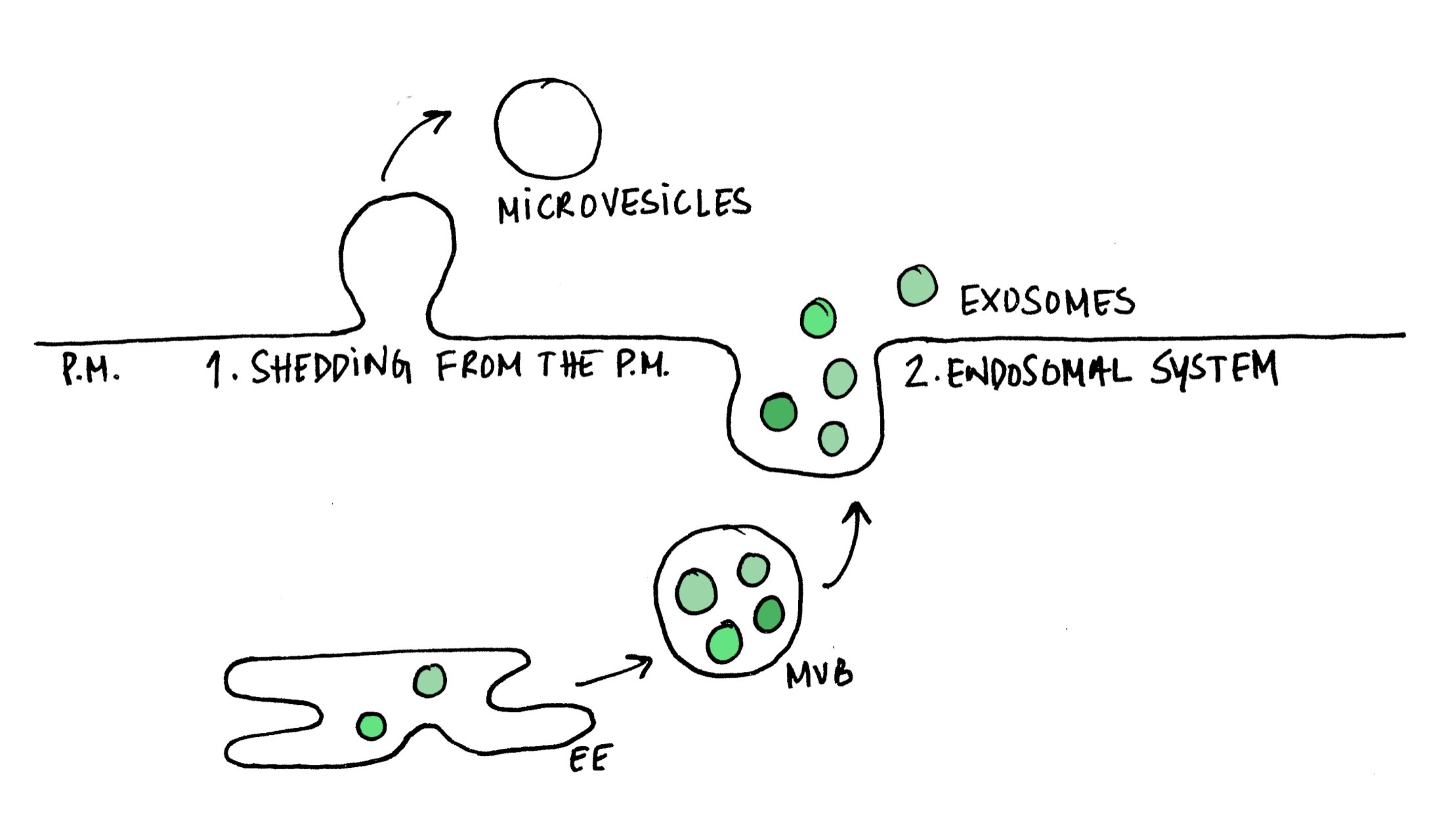
van Niel G, D'Angelo G and Raposo G. Shedding light on the cell biology of extracellular vesicles. Nat Rev Mol Cell Biol 19, 213-228 (2018). https://doi.org/10.1038/nrm.2017.125
Kowal J, Arras G, Colombo M, Jouve M, Morath JP, Primdal-Bengtson B, Dingli F, Loew D, Tkach M and Théry C. Proteomic comparison defines novel markers to characterize heterogeneous populations of extracellular vesicle subtypes. Proc Natl Acad Sci USA 113(8), E968-E977 (2016). https://doi.org/10.1073/pnas.152123011
Lo Cicero A, Delevoye C, Gilles-Marsens F, Loew D, Dingli F, Guere C, Andre N, Vie K, van Niel G and Raposo G. Exosomes released by keratinocytes modulate melanocyte pigmentation. Nat Commun 6, 7506 (2015). https://doi.org/10.1038/ncomms8506
Primary keratinocytes secrete exosomes that modulate the pigmentation status of melanocytes. This manuscript shows for the first time that exosomes are essential in skin homeostasis and the maintenance of pigmentation. Exosomes were shown to induce transcription factors and activate genes affecting melanosome structure and function. These effects are attributed to particular miRNAs associated with the exosomes. This study opened a pathway to develop cosmetic treatments to modulate pigmentation.
Ostrowski M, Carmo NB, Krumeich S, Fanget I, Raposo G, Savina A, Moita CF, Schauer K, Hume AN, Freitas RP, Goud B, Benaroch P, Hacohen N, Fukuda M, Desnos C, Seabra MC, Darchen F, Amigorena S, Moita LF and Thery C. Rab27a and Rab27b control different steps of the exosome secretion pathway. Nature Cell Biology 12, 19–30 (2010). https://doi.org/10.1038/ncb2000
Fevrier B, Vilette D, Archer F, Loew D, Faigle W, Vidal M, Laude H and Raposo G. Cells release prions in association with exosomes. Proc Natl Acad Sci U S A 101, 9683-9688 (2004). https://doi.org/10.1073/pnas.030841310
Prions can be secreted from cells in association with exosomes. This manuscript shows for the first time that exosomes carrying infectious Prions are able to induce neurodegenerative disease in mouse models. This study provided the underpinnings for a wide range of studies, from basic to applied, to further understand the pathophysiology of neurodegenerative diseases and the role of extracellular vesicles as purveyors of the infection cycle.
Raposo G, Nijman HW, Stoorvogel W, Leijendekker R, Harding CV, Melief CJ and Geuze HJ. B lymphocytes secrete antigen-presenting vesicles. J Exp Med 183, 1161-1172 (1996). https://doi.org/10.1084/jem.183.3.1161
This is the first study to reveal that exosomes operate as signaling entities. This manuscript shows that cells from the immune system, such as B lymphocytes, secrete endosome-derived exosomes. Importantly, exosomes derived from both human and murine B lymphocytes induced antigen-specific MHC class II-restricted T cell responses, suggesting a role for exosomes in antigen presentation in vivo.
Ilustrations by Ana Soares
Extracelular Vesicles: Microvesicle and Exosomes
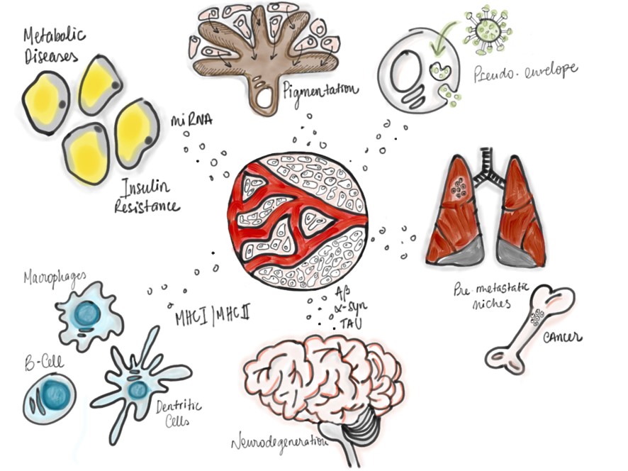
Extracelular Vesicles are involved in many physiological and disease contexts.
NOVA Medical School - LisboaCampo Mártires da Pátria, 1301169-056 LisboaPORTUGAL
NOVA Medical School - CarcavelosRua de Luanda 166,2775-233 ParedePORTUGAL
GeneralTel.: +351 218 803 000
Contacts
Compliment, suggestion or complaint
WHISTLEBLOWER PORTAL
Students Association
News and Events
Awards and Honours
IT Support
IT Resources
Whistleblower Portal
Plano de Recuperação e Resiliência (PRR) Programa Lisboa 2030